Select Language:
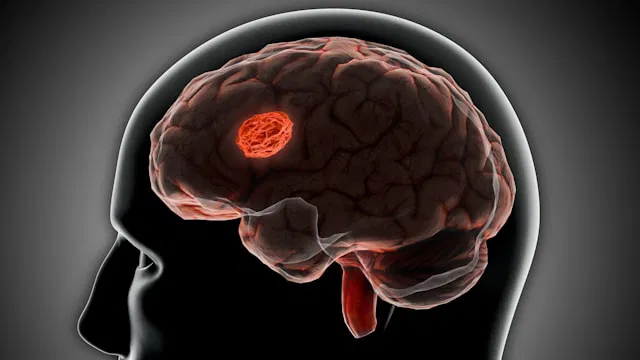
A stroke is a critical medical event that occurs when blood flow to a part of the brain is blocked or reduced. This interruption can cause damage to brain cells, leading to difficulties with movement, speech, and cognition.
Many stroke survivors face persistent physical challenges, particularly weakness or paralysis on one side of the body.
For years, researchers have been exploring how the brain recovers after a stroke. A recent study published in The Lancet Digital Health offers new insights. It suggests that the brain may adapt in ways that scientists hadn’t fully understood before.
The study was conducted by researchers from the University of Southern California, with collaboration from an international team through the global ENIGMA network. They examined brain scans from more than 500 stroke survivors across 34 research centers in eight different countries.
The scientists utilized sophisticated artificial intelligence algorithms to analyze MRI scans. They trained models using tens of thousands of brain images to estimate the “age” of different brain regions. It’s important to note that this “brain age” isn’t related to the person’s actual age but reflects how healthy or deteriorated that brain region appears.
The findings showed that areas of the brain affected by stroke tend to age more rapidly. This isn’t entirely surprising, as injury can weaken brain tissue. What was unexpected, however, was that the healthy side of the brain sometimes appeared biologically younger than expected.
This “youthful” appearance was most evident in patients with more severe movement impairments. Even months after the stroke, these individuals displayed signs that their undamaged brain regions had undergone changes making them look biologically younger.
Scientists think this might be a sign that the brain is trying to adapt. When one part of the brain sustains damage, other areas may step in to compensate. This process is called neuroplasticity—the brain’s remarkable ability to rewire itself.
The study identified the involvement of a particular network called the frontoparietal network, which assists with movement, attention, and coordination. In patients with severe impairments, this network on the healthy side of the brain showed a more “youthful” pattern, indicating adaptive changes.
It’s important to understand that this doesn’t mean the brain has fully healed. Rather, it indicates that the brain is exerting extra effort to regain function, reorganizing itself to help patients move more effectively and perform daily activities.
An advantage of this study was its large sample size. By analyzing data from hundreds of participants worldwide, researchers could identify patterns that smaller studies might miss. The use of AI also enabled a deeper understanding of complex brain data in ways that weren’t possible before.
That said, the research has limitations. It was based on a single snapshot in time and did not follow patients over years. As a result, it can’t fully explain how these brain changes evolve during recovery.
Ultimately, this research offers fresh perspectives on how the brain reacts to injury. It shows that recovery involves more than just repairing damage; it also includes the brain’s capacity to adapt and reorganize. These insights could lead to more personalized rehab strategies tailored to each individual’s brain changes in the future.
If you’re interested in stroke-related health tips, check out studies on diets rich in flavonoids that may lower stroke risk and the MIND diet, which might slow cognitive decline post-stroke.
For additional health insights, explore recent studies on antioxidants that could help prevent dementia, and how drinking tea or coffee may reduce the risk of stroke and cognitive issues in articles like tea and coffee’s potential benefits.
Source: University of Southern California.